

Health technology
DeepCancer — Explainable AI for Breast Histopathology
Pathology AI for computational and digital pathology: hybrid models, Grad-CAM explainability, and a research stack built for measurable review—not black-box classification.
- Research
- Murat Onur Kaderoğlu · Emre Şatır
- Project type
- Applied AI / research prototype
- Field
- Digital pathology · computational pathology · breast histopathology
- Core technology
- Swin Transformer + ConvNeXt · Grad-CAM
- Status
- Working research prototype
- Use scope
- Research and education
- Research publication
- Journal of Artificial Intelligence with Applications · 2025
Published test results
Reported binary benign / malignant classification results on the BreaKHis dataset; these are not clinical-use results.
Problem and research context
Histopathology is not only a classification problem: specialists also need to examine which image regions affect a model output. DeepCancer brings benign / malignant classification, probabilistic output, and Grad-CAM visual explanation into one research workflow.
What the research system provides
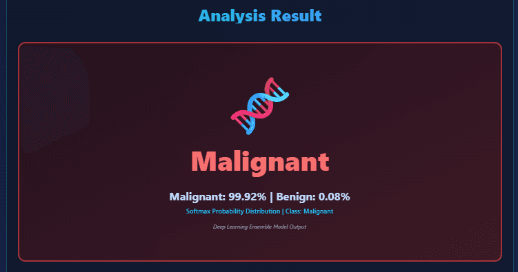
The web workflow connects histopathology image intake, image preparation and validation, hybrid Swin Transformer + ConvNeXt analysis, probabilistic benign / malignant classification, a Grad-CAM focus map, and expert review.
Why a hybrid architecture?
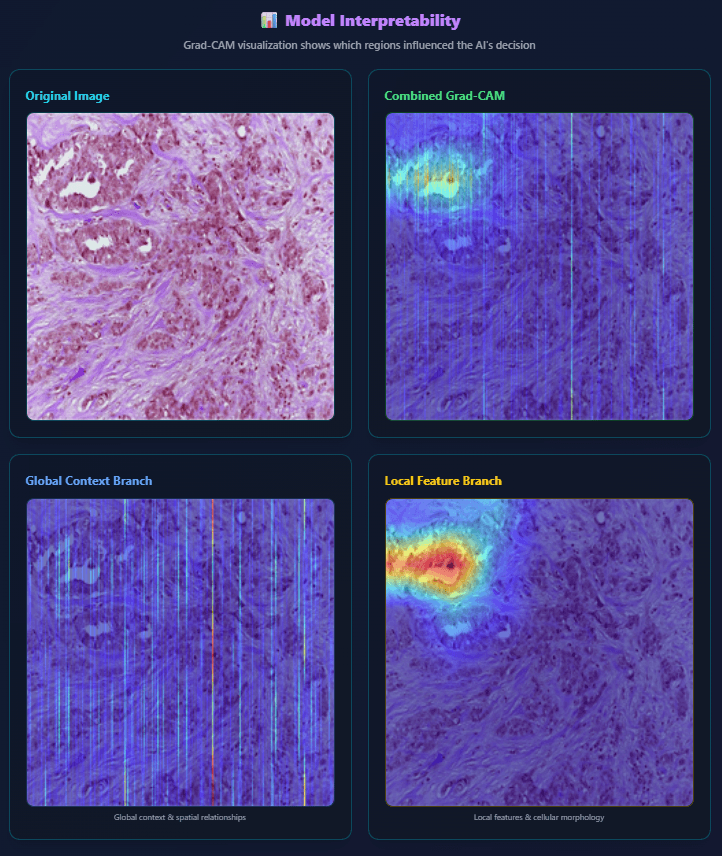
ConvNeXt supports representation of local and spatial patterns, while Swin Transformer models wider contextual relationships through window-based attention. The aim is to use the complementary representation strengths in one classification task.
Explainability
Grad-CAM presents image regions affecting a prediction as a focus map. It does not prove that a model decision is correct and does not replace pathological interpretation; it supplies additional context for research review.
Research and validation
The published study uses 7,909 H&E images from 82 patients in the open-access BreaKHis dataset, at 40×, 100×, 200×, and 400× magnification. Images are resized to 224 × 224 and evaluated with the hybrid architecture.
From research to a working system
The prototype takes a suitable image, produces a probabilistic output with the hybrid model, visualizes focus regions with Grad-CAM, and presents classification and visual explanation together for expert review.
Current limitations
DeepCancer is not a medical device, is not FDA- or CE-approved for clinical diagnosis, and is not offered for diagnostic or treatment decisions. Results belong to experimental BreaKHis evaluation; external clinical validation and relevant ethical, technical, and regulatory processes are required.
Screens from the working research prototype
These screens are from the research prototype. Probabilistic classification and Grad-CAM visualisation are not used for clinical diagnosis or treatment decisions.
Primary sources
From research to a working AI system
TankDev turns image analysis, bespoke AI models, and explainable AI approaches from research prototypes into working software systems.
Explore Applied AI